RetInSight GA Monitor
The RetInSight GA Monitor for Geographic Atrophy (GA)
AI-based segmentation in clinical practice: Making the invisible visible

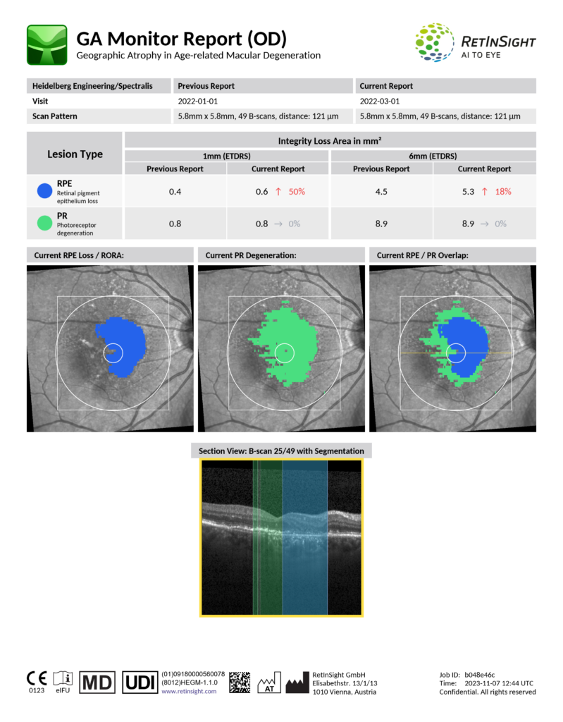
Sample of a RetInSight GA Monitor PDF Report
Key features of the RetInSight GA Monitor
- AI-based Segmentation: Advanced AI algorithms enable precise segmentation of GA areas
- Disease Activity Assessment: Measure and visualize disease activity and therapeutic efficacy across all aspects, including
- Precise and Plausible Disease Monitoring: Visualize and track disease progression, allowing for precise and timely interventions
- Regulatory Compliance: The RetInSight GA Monitor is the first and only AI-based software fulfilling all MDR 2017/745 requirements for Class IIa medical devices as Clinical Decision Support Systems (CDSS) for the monitoring of patients with GA
Sample of a RetInSight GA Monitor PDF Report
Why the RetInSight GA Monitor is a game-changer in GA management
The RetInSight GA Monitor has been thoughtfully crafted to address the unique challenges of GA diagnosis and progression tracking:
- Unique photoreceptor visualization: Be at the forefront of GA management with the unique ability to measure photoreceptor degeneration, the primary sign of GA and the essence of vision1
- Identify progression: Distinguish slow vs. fast progressors at first presentation and track the speed of development of RPE and PR integrity loss in each patient and over time, enabling personalized treatment and a realistic assessment of therapeutic benefit3, 4, 5
- Efficient workflow: Integrate GA management seamlessly into your current OCT routine, streamlining workflow and enhancing efficiency with a mouse click
- Quality control: Replace time-consuming and error-prone human image analysis with fast, reproducible, and quantifiable data supporting informed therapeutic decisions
- Patient communication: Foster patient compliance by delivering customized reports also to patients, ideally reducing the risk of over- and under-treatment and increasing trust with self-explanatory, personalized reports
Empowering GA management through visualization and quantification of PR degeneration and RPE loss on standard OCT images
As a trusted leader in retinal care, RetInSight invites you to explore the GA Monitor and experience the next generation of GA management. Our innovative technology will empower you to make informed treatment decisions based on precise measurements and visualizations of PR and RPE loss, all while remaining committed to preserving your patients’ vision throughout their lifetime.
The RetInSight GA Monitor is the first and only AI-based software fulfilling all MDR 2017/745 requirements for Class IIa medical devices as Clinical Decision Support Systems (CDSS) for the monitoring of patients with GA.
For more information and a demo of the RetInSight GA Monitor, please get in touch with our team. Together, let’s revolutionize the way we manage and treat geographic atrophy.
References
- Julia Mai et al. | Comparison of Fundus Autofluorescence Versus Optical Coherence Tomography–based Evaluation of the Therapeutic Response to Pegcetacoplan in Geographic Atrophy
- Sophie Riedl et al. | The Effect of Pegcetacoplan Treatment on Photoreceptor Maintenance in Geographic Atrophy Monitored by Artificial Intelligence – Based OCT Analysis
- Wolf-Dieter Vogl et al. | Predicting Topographic Disease Progression and Treatment Response of Pegcetacoplan in Geographic Atrophy Quantified by Deep Learning
- Ursula Schmidt-Erfurth et al. | Monitoring der Progression von geografischer Atrophie in der optischen Kohärenztomographie
- Ursula Schmidt-Erfurth et al. | Disease activity and therapeutic response to pegcetacoplan for geographic atrophy identified by deep learning-based analysis of OCT
FAQs
What does the RetInSight GA Monitor measure on OCT scans?
The GA Monitor provides automated detection, segmentation, and quantification of areas consistent with geographic atrophy (GA) based on OCT image data, and in patients diagnosed with GA.
It is intended for visualization and quantification of Retinal Pigment Epithelium (RPE) Loss and Photoreceptor (PR) Degeneration. The PR/RPE overlap zone, provides clinicians with additional information for evaluating structural changes over time.
How does the GA Monitor address the limitations of FAF in GA assessment?
Fundus autofluorescence (FAF) is commonly used to assess RPE loss in GA, but it has limitations:
- FAF primarily visualizes RPE loss, not photoreceptor degeneration, which is also relevant in GA.
- FAF requires additional imaging equipment that not all clinics have.
- FAF can be uncomfortable for patients due to the intense light exposure.
The GA Monitor uses routine OCT imaging, which is already part of most clinics’ workflows, and provides information on both RPE and photoreceptor structure derived from OCT data.
How can automated GA quantification support clinical decision‑making?
The GA Monitor provides objective, quantitative OCT‑based information that clinicians may use as additional input when documenting GA progression or reviewing changes between visits.
Does the GA Monitor integrate with existing OCT devices and clinical workflows?
The GA Monitor currently works exclusively with the Heidelberg Spectralis OCT. This is intentional.
Different OCT devices vary significantly in image quality, resolution, noise, and acquisition protocols. If you compare a Spectralis scan with one from another device, the difference is clear, and these differences directly influence how reliably an algorithm can detect and quantify biomarkers.
An algorithm optimized and validated on Spectralis images will not necessarily perform the same way on images from another OCT.
To avoid unreliable or inconsistent results, RetInSight does not use a “one‑algorithm‑fits‑all‑devices” approach. Each OCT platform requires its own dedicated integration and validation. Support for additional devices is planned and will be released once validated for that specific hardware.
Why isn’t the GA Monitor device‑agnostic?
Because OCT devices are not created equal.
They differ in scan quality, resolution, noise characteristics, number of B‑scans, scanning speed, and acquisition consistency. These differences mean that AI algorithms trained on one device will not automatically deliver the same performance on another.
Particularly for layers relevant in GA such as photoreceptors, studies show that Spectralis OCT offers high image consistency.
The GA Monitor algorithms were trained and validated on Spectralis data, ensuring reliable processing of the associated image characteristics.
Support for additional devices will follow after device‑specific validation.
How frequently can GA be monitored using the GA Monitor?
The GA Monitor can analyze GA-related biomarkers whenever a Spectralis OCT scan is acquired.
Monitoring intervals depend on individual clinic workflows and clinician judgment for patients diagnosed with GA.
Is the GA Monitor supported by clinical evidence?
The underlying algorithms have undergone technical and clinical performance evaluations as required under MDR, UK MDR, TGA, and Medsafe.
Details on validation studies can be provided upon request.
How can the GA Monitor support standardization across clinics or clinical teams?
By using the same algorithmic criteria for every Spectralis OCT scan, the GA Monitor delivers consistent, quantitative results that support standardized documentation within multi‑clinician or multi‑site environments.
Can the GA Monitor be used together with other RetInSight tools in clinical routine?
Yes. The GA Monitor is part of the RetInSight Clinical Routine Suite and can be used alongside other AI tools, such as the Fluid Monitor.
Each tool provides separate, complementary quantitative information based on Spectralis OCT imaging.
What additional insights does photoreceptor (PR) analysis provide in GA monitoring?
Photoreceptor degeneration is an important structural feature in GA.
The GA Monitor provides automated quantification of PR layer changes from OCT scans, offering clinicians additional information on structural integrity beyond the RPE layer.
This may support a more comprehensive view of structural changes in patients diagnosed with GA.
Why is OCT‑based GA monitoring valuable for clinics that do not use FAF?
OCT is widely available in routine care, and most clinics already use it for AMD assessments.
With the GA Monitor:
Clinics can evaluate GA‑related structural changes without additionally requiring FAF.
They receive quantitative OCT‑based metrics that provide additional information about both RPE and photoreceptor layers.
They avoid the additional patient discomfort associated with FAF scans.